An Integrated Approach to Ultrasound Imaging in Medicine and Biology
Co-Editor-in-Chief, BIO Integration
Published Online: October 16 2020
Cite this paper:
Pingtong Huang. An Integrated Approach to Ultrasound Imaging in Medicine and Biology. BIO Integration 2020; 1(3): 105–109.
DOI: 10.15212/bioi-2020-0036. Available at: https://bio-integration.org/
Download citation 
© 2020 The Authors. This is an open access article distributed under the terms of the Creative Commons Attribution License (https://creativecommons.org/licenses/by/4.0/). See https://bio-integration.org/copyright-and-permissions/
Ultrasound (US) imaging is developing rapidly as it enhances the specificity and sensitivity of medical imaging. The disadvantages pertaining to US imaging include dependence on the operator’s skill and equipment, the limited acoustic window in certain patients [chronic obstructive pulmonary disease (COPD), obesity], limitations when used for examinations of gas-containing body regions (such as the lungs and the digestive system), and the relative inability of US to penetrate bone. Many of these problems can only be solved through multidisciplinary integration and technical cooperation [1]. Herein, we examine the present status and provide an outlook on the progress of US imaging and therapeutic applications in the near future.
US has been extensively applied in the diagnostic process of clinical diseases because of its advantages of non-invasiveness, non-radiation, convenience, and low cost. As opposed to a mechanical wave, US can enter the human body non-invasively. The acoustic impedances of various tissues and organs of the human body are different, which in turn causes ultrasonic waves to generate echoes during their propagation through human tissue. The echo carries information regarding the disparities in the acoustic impedance attributed to the tissues. Obtaining this echo information can produce structural images of tissues and organs, thereby helping clinicians to diagnose diseases.
Current clinical US imaging
B-mode imaging
The primary functions of US scanners are to render real-time images of underlying tissue [2]. Specifically, B-mode imaging is utilized for imaging the majority of organs in the human body including, through the development of new techniques, the detection of lung and bone lesions.
US Doppler imaging
The movement of red blood cells (RBCs), which represents the flow of blood, can dissipate weak echoes of US and is detectable via US Doppler imaging [3]. US Doppler imaging generates vascular images demonstrating either the energy or the velocity of RBC echoes.
Three-dimensional US imaging
The modality of three-dimensional (3D) US imaging includes static 3D imaging and dynamic 3D imaging. The latter is a global imaging method, which is used to reconstruct a 3D image of the real-time activity of the region of interest. One of the representations is four-dimensional echocardiography.
Contrast-enhanced US imaging
Contrast-enhanced US imaging is dependent on the administration of US contrast agents (UCAs) for the enhancement of the ultrasonic signals in the detection and improvement in the quality of US images. Contrast-enhanced US imaging has been commonly utilized in clinical practices for the delineation of suspicious lesions in multiple organs (such as the liver) as well as identification of abnormalities in the cardiovascular system [4]. Gas-filled microbubbles provide enhancement to contrast-enhanced US imaging. The flow of blood distributes these microbubbles throughout the body, which generates non-linear signals with harmonic frequencies and linear backscatter under varying acoustic pressures [5]. Contrast-enhanced images are conducive to the dynamic surveillance of organs and blood vessels.
Emerging US imaging
Elastography imaging
Elastography imaging has made great progress in the past decade for imaging of the mechanical characteristics of tissue. Elastography imaging mainly includes: quasi-static elastography (QE) [6], transient elastography (TE) [7], acoustic radiation force imaging (ARFI) [8], as well as shear wave imaging (SWI) [9], etc. SWI enables the clinical imaging of the shear modulus of tissues or the shear-wave speed [10]. These techniques demonstrate its main clinical application in the diagnosis of liver fibrosis [11] and assessment of tumors in the breast [12], or thyroid gland [13]. And now, its application has been extended to the diagnosis of prostatic lesions, kidney diseases, etc. Additionally, within heterogeneous media, the observed wave speed can be calculated not only from the properties of the tissue but also by its boundaries, which can steer the waves and alter the observed speed.
US molecular imaging
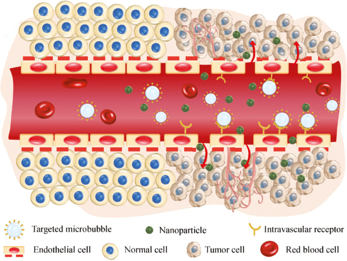
The field of US molecular imaging is an amalgamation of molecular imaging, US medicine, nanomedicine, and material science with the exclusive benefits of precision, non-invasiveness, and repeatability, which enhances the specificity and sensitivity of US diagnosis [14, 15]. Active targeting modification allows UCAs to aggregate at a specific site by the binding of peptides or ligands onto the surface, which is a common approach within the research of US molecular imaging [16]. Apart from the ligand alteration of the contrast agent to accomplish active targeting, a broad range of targeted UCAs have been developed by researchers for various applications to accommodate to the intricate internal environment of the human body, which subsequently enhances the applicational and functional value of the contrast agent. Therefore, nanoscale UCAs can be accumulated outside blood vessels due to the enhanced permeability and retention (EPR) effect to accomplish tumor tissue imaging (Figure 1). Targeting UCAs have been employed in the diagnosis and management of numerous diseases, such as angiogenesis [17, 18], inflammation [19, 20], and thrombosis [21].
Figure 1 Ligand-coupled Microbubbles (MBs) actively binds to the intravascular receptors of the endothelium of tumors in addition to the penetration of nanoparticles into tumor issue through the enhanced permeation and retention (EPR) effect.
Ultrafast imaging
Ultrafast US imaging technology is based on the plane wave US imaging method for tissue imaging. In one imaging procedure, all transducer array elements are excited for pulse transmission, and the echo data of all array elements are received and processed at the same time. The transducer only needs one transmission and reception to obtain US images in the sound field, so it can achieve US data acquisition and imaging at a frame rate much higher than traditional focused US imaging [22]. At the same time, the new US imaging algorithm is able to substantially enhance the sensitivity of imaging of blood flow, provide technical support for micro blood flow imaging, and also expand the application of brain US imaging technology [23].
Super-resolution US imaging
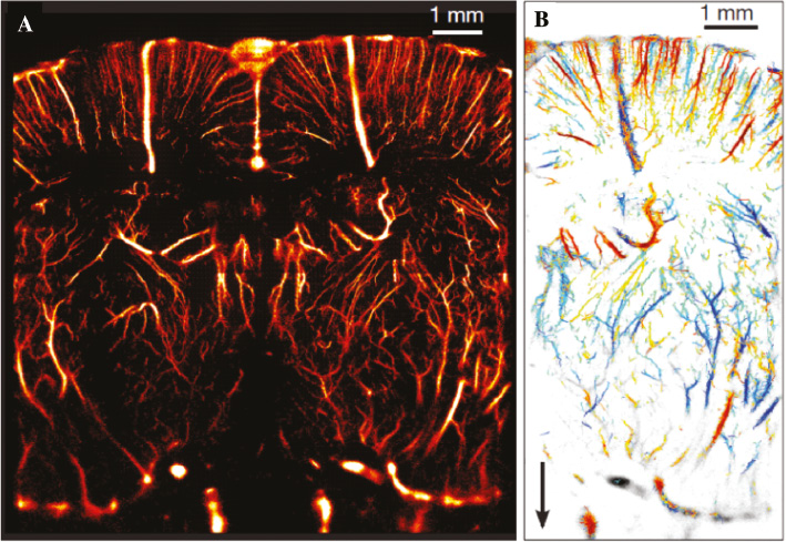
High-resolution imaging has consistently been an important research direction in biomedical imaging. However, due to the problem of the imaging diffraction limit, the resolution of traditional US imaging will not exceed the half-wavelength of the US used. In recent years, it has been found that US can also achieve super-resolution US imaging by using special media to break the limitation of the diffraction limit. In 2015, Tanter’s team made an important breakthrough in obtaining super-resolution brain blood flow imaging by using ultra-fast US imaging and super-resolution US imaging of microbubble tracing (Figure 2) [24]. The achieved resolution was more than 10 μm [25], which breaks through the diffraction limit of traditional US imaging, and improves the resolution of blood flow ultrasonographic imaging by orders of magnitudes, which greatly expands the applicational space of US imaging [26]. It is worth noting that high-resolution acoustic imaging technology is inseparable from the development of high-quality new piezoelectric materials or acoustic materials, reinforcing that the fusion of different technologies is the cornerstone of development.
Figure 2 Mouse brain super-resolution imaging depicting minute vascular structures in addition to corresponding blood flow velocities. (A) Micro US localization microscopy (μULM) conducted through a thinned skull in the coronal section, bregma −1.5 mm, which delivers a resolution of 10 μm × 8 μm in depth and lateral direction, respectively; (B). In-plane velocity maps from parts of the vessels in (A) [25].
Photoacoustic imaging
Photoacoustic (PA) imaging is an emerging imaging technology utilizing a non-invasive, non-ionizing, real-time imaging method which is able to visualize the optical absorption properties of tissue with great special resolution and at a reasonable depth. With the swiftly developing targeted nano/microbubble agents, in vivo molecular imaging is achievable via PA, which facilitates further cellular and molecular assessment of tissue, which has been employed in wound healing, cancer imaging, gene expression, disorders of the brain as well as the acquisition of functional information about the tissue.
Opportunities and challenges
In recent years, the bottleneck on clinical US imaging, for example, the lack of experience and skill of the examining physician exerted great influence on the outcome of the reliability of their diagnosis. However, some of these bottlenecks can be solved by integrating other interdisciplinary such as artificial intelligence technology. Implementation of true 3D imaging, automated image analysis, and image post-processing tools, and further development of standardization of scanning protocols and data files can address these limitations. Solving these problems may also stimulate emerging multidisciplinary intersections and set the stage for improved clinical diagnosis and therapeutic applications.
On the other hand, functional US contrast agents have not achieved realization from bench to bedside. BR55, taken as an example here, is a particular clinical-grade UCA which demonstrates targeted binding to vascular endothelial growth factor receptor 2 (VEGFR2), and exhibits encouraging prospects for the imaging of tumors in a multitude of preclinical models. Animal experiments conducted by Hackl et al. [27] established that the implementation of high-resolution BR55 technology was able to achieve sensitive and non-invasive discovery of early micrometastases of tumors, exhibiting the prospect for early diagnosis of tumors. Since then, additional in vivo and in vitro BR55 has begun to be implemented in clinical trials. For example, Smeenge et al. [28] studied the safety and possibility of utilizing contrast agents for the identification of prostate cancer in humans. Twenty-four patients who were diagnosed with prostate cancer by biopsy were selected, and BR55 contrast-mediated imaging was conducted with clinical low-acoustic intensive US. As this is the first in-human pioneer clinical trial, the imaging procedures and the dose of contrast agent were thoroughly considered. Results demonstrated that contrast agent at doses of 0.03 ml/kg and 0.05 ml/kg were adequate to acquire an enhanced contrast image for 30 min. No significant side effects were observed, indicating after the safety of this procedure.
One of the essential regulators of neo-angiogenesis in cancer is kinase-insert domain receptor (KDR). Utilizing a clinical-grade KDR-targeted contrast microbubble (MBKDR), the first in-human clinical trial on US molecular imaging in patients with ovarian and breast lesions was conducted by Willmann et al. [29]. Twenty-one women with focal breast lesions and 24 women with focal ovarian lesions received intravenous injection of MBKDR. Results indicated that KDR-targeted USMI signal demonstrated good matching with KDR expression using immunohistochemistry (IHC), with the advantage of immediate tracing following intravenous injection. No patients reported adverse events, and there were no aberrations or substantial changes of trends in electrocardiograms (ECGs), vital signs, or measured laboratory tests. These results indicated that US molecular imaging is feasible, safe, and allows non-invasive clinical assessment of the expression of KDR in patients.
Future outlook
US imaging is an important method for clinical disease screening, early diagnosis, and non-invasive guidance. The development and research pertaining to UCAs for US molecular imaging portrays the essence of the advancement of US targeted technologies and the driving force for the progression of US molecular imaging. The principal path of existing US molecular imaging would be pre-clinical experimental research evolving to clinical application. Additionally, the clinical implementation of new methods and knowledge in the field of US molecular imaging to assist patients is an obstacle for future research of US molecular imaging.
Inherent advantages pertaining US is that it applies basic physical stimulation to living tissue. The fields of molecular US and US genetics seek to link these physical forces with the functions of biomolecules and cells to achieve precise control of cells. In addition, genetic engineering allows biomolecules to interact with US waves. For example, the thermal effect or mechanical effect of US can be used to control temperature-sensitive and mechanically-sensitive bacteria. Although this field is still in its infancy, it has opened the door to precise US control. Concurrently, the utilization of US-mediated microbubble cavitation to augment the movement of genes, drugs, and antibodies across the vascular endothelial barrier and realize intracellular delivery has created new opportunities for applications in biology and clinical transformation. Furthermore, sonodynamic therapy (SDT) has surfaced as a favorable option for the minimally-invasive treatment of solid cancers. The compounded utilization of US and a sonosensitizer drug to produce cytotoxic reactive oxygen species (ROS) in and around neoplastic cells forms the basis of SDT.
In the near future, it will be possible that the rise in cross-disciplinary and interdisciplinary cooperation will continue to promote the advancement of US theranostic techniques with additional accomplishments in clinical trials and basic research which could have extensive benefits to human health.
Reference
- Saw PE, Jiang S. The significance of interdisciplinary integration in academic research and application. BIO Integration 2020;1:2-5. [DOI: 10.15212/bioi-2020-0005]
- Powers J, Kremkau F. Medical ultrasound systems. Interface Focus 2011;1:477-89. [PMID: 22866226 DOI: 10.1098/rsfs.2011.0027]
- Evans DH, Jensen JA, Nielsen MB. Ultrasonic colour Doppler imaging. Interface Focus 2011;1:490-502. [PMID: 22866227 DOI: 10.1098/rsfs.2011.0017]
- Claudon M, Cosgrove D, Albrecht T, Bolondi L, Bosio M, et al. Guidelines and good clinical practice recommendations for contrast enhanced ultrasound (CEUS) – Update 2008. Ultraschall in Der Medizin 2008;29:28-44. [PMID: 18270887 DOI: 10.1055/s-2007-963785]
- Son S, Min HS, You DG, Kim BS, Kwon IC. Echogenic nanoparticles for ultrasound technologies: evolution from diagnostic imaging modality to multimodal theranostic agent. Nano Today 2014;9:525-40. [DOI: 10.1016/j.nantod.2014.06.002]
- Ophir J, Cespedes I, Ponnekanti H, Yazdi Y, Li X. Elastography – A quantitative method for imaging the elasticity of biological tissues. Ultrason Imaging 1991;13:111-34. [PMID: 1858217 DOI: 10.1177/016173469101300201]
- Catheline S, Thomas JL, Wu F, Fink MA. Diffraction field of a low frequency vibrator in soft tissues using transient elastography. IEEE Trans Ultrason Ferroelectr Freq Control 1999;46:1013-9. [PMID: 18238506 DOI: 10.1109/58.775668]
- Nightingale K, Soo MS, Nightingale R, Trahey G. Acoustic radiation force impulse imaging: in vivo demonstration of clinical feasibility. Ultrasound Med Biol 2002;28:227-35. [PMID: 11937286 DOI: 10.1016/s0301-5629(01)00499-9]
- Bercoff J, Tanter M, Fink M. Supersonic shear imaging: a new technique for soft tissue elasticity mapping. IEEE Trans Ultrason Ferroelectr Freq Control 2004;51:396-409. [PMID: 15139541 DOI: 10.1109/tuffc.2004.1295425]
- Lee SH, Chang JM, Kim WH, Bae MS, Seo M, et al. Added value of shear-wave elastography for evaluation of breast masses detected with screening US imaging. Radiology 2014;273:61-9. [PMID: 24955927 DOI: 10.1148/radiol.14132443]
- Wang C-Z, Zheng J, Huang Z-P, Xiao Y, Song D, et al. Influence of measurement depth on the stiffness assessment of healthy liver with real-time shear wave elastography. Ultrasound Med Biol 2014;40:461-9. [PMID: 24361224 DOI: 10.1016/j.ultrasmedbio.2013.10.021]
- Xiao Y, Zeng J, Niu L, Zeng Q, Wu T, et al. Computer-aided diagnosis based on quantitative elastographic features with supersonic shear wave imaging. Ultrasound Med Biol 2014;40:275-86. [PMID: 24268454 DOI: 10.1016/j.ultrasmedbio.2013.09.032]
- Bhatia KSS, Lam ACL, Pang SWA, Wang D, Ahuja AT. Feasibility study of texture analysis using ultrasound shear wave elastography to predict malignancy in thyroid nodules. Ultrasound Med Biol 2016;42:1671-80. [PMID: 27126245 DOI: 10.1016/j.ultrasmedbio.2016.01.013]
- Gao Y, Hernandez C, Yuan H-X, Lilly J, Kota P, et al. Ultrasound molecular imaging of ovarian cancer with CA-125 targeted nanobubble contrast agents. Nanomed-Nanotechnol Biol Med 2017;13: 2159-68. [PMID: 28603079 DOI: 10.1016/j.nano.2017.06.001]
- Wang S, Hossack JA, Klibanov AL. Targeting of microbubbles: contrast agents for ultrasound molecular imaging. J Drug Target 2018;26: 420-34. [PMID: 29258335 DOI: 10.1080/1061186X.2017.1419362]
- Kaneko OF, Willmann JK. Ultrasound for molecular imaging and therapy in cancer. Quant Imaging Med Surg 2012;2:87-97. [PMID: 23061039 DOI: 10.3978/j.issn.2223-4292.2012.06.06]
- Wang J, Qin B, Chen X, Wagner WR, Villanueva FS. Ultrasound molecular imaging of angiogenesis using vascular endothelial growth factor-conjugated microbubbles. Mol Pharm 2017;14:781-90. [PMID: 28165246 DOI: 10.1021/acs.molpharmaceut.6b01033]
- Yan F, Xu X, Chen Y, Deng Z, Liu H, et al. A lipopeptide-based alpha V beta(3) integrin-targeted ultrasound contrast agent for molecular imaging of tumor angiogenesis. Ultrasound Med Biol 2015;41:2765-73. [PMID: 26166460 DOI: 10.1016/j.ultrasmedbio.2015.05.023]
- Machtaler S, Knieling F, Luong R, Tian L, Willmann JK. Assessment of inflammation in an acute on chronic model of inflammatory bowel disease with ultrasound molecular imaging. Theranostics 2015;5:1175-86. [PMID: 26379784 DOI: 10.7150/thno.13048]
- Suzuki J-i, Ogawa M, Takayama K, Taniyama Y, Morishita R, et al. Ultrasound-microbubble-mediated intercellular adhesion molecule-1 small interfering ribonucleic acid transfection attenuates neointimal formation after arterial injury in mice. J Am Coll Cardiol 2010;55:904-13. [PMID: 20185042 DOI: 10.1016/j.jacc.2009.09.054]
- Wang X, Gkanatsas Y, Palasubramaniam J, Hohmann JD, Chen YC, et al. Thrombus-targeted theranostic microbubbles: a new technology towards concurrent rapid ultrasound diagnosis and bleeding-free fibrinolytic treatment of thrombosis. Theranostics. 2016;6:726-38. [PMID: 27022419 DOI: 10.7150/thno.14514]
- Bourdeau RW, Lee-Gosselin A, Lakshmanan A, Farhadi A, Kumar SR, et al. Acoustic reporter genes for noninvasive imaging of microorganisms in mammalian hosts. Nature 2018;553:86-90. [PMID: 29300010 DOI: 10.1038/nature25021].
- Tanter M, Fink M. Ultrafast imaging in biomedical ultrasound. IEEE Trans Ultrason Ferroelectr Freq Control 2014;61:102-19. [PMID: 24402899 DOI: 10.1109/TUFFC.2014.6689779]
- Desailly Y, Couture O, Fink M, Tanter M. Sono-activated ultrasound localization microscopy. Appl Phys Lett 2013;103:174107. [DOI: 10.1063/1.4826597]
- Errico C, Pierre J, Pezet S, Desailly Y, Lenkei Z, et al. Ultrafast ultrasound localization microscopy for deep super-resolution vascular imaging. Nature 2015;527:499-502. [PMID: 26607546 DOI: 10.1038/nature16066]
- Couture O, Hingot V, Heiles B, Muleki-Seya P, Tanter M. Ultrasound localization microscopy and super-resolution: a state of the art. IEEE Trans Ultrason Ferroelectr Freq Control 2018;65: 1304-20. [PMID: 29994673 DOI: 10.1109/TUFFC.2018.2850811]
- Hackl C, Schacherer D, Anders M, Wiedemann LM, Mohr A, et al. Improved detection of preclinical colorectal liver metastases by high resolution ultrasound including molecular ultrasound imaging using the targeted contrast agent BR55. Ultraschall Der Medizin 2016;37:290-6. [PMID: 27112624 DOI: 10.1055/s-0041-111838]
- Smeenge M, Tranquart F, Mannaerts CK, de Reijke TM, van de Vijver MJ, et al. First-in-human ultrasound molecular imaging with a VEGFR2-specific ultrasound molecular contrast agent (BR55) in prostate cancer. A safety and feasibility pilot study. Investigative Radiol 2017;52:419-27. [PMID: 28257340 DOI: 10.1097/RLI.0000000000000362]
- Willmann JK, Bonomo L, Testa AC, Rinaldi P, Rindi G, et al. Ultrasound molecular imaging with BR55 in patients with breast and ovarian lesions: first-in-human results. J Clin Oncol 2017;35:2133-40. [PMID: 28291391 DOI: 10.1200/JCO.2016.70.8594]